
Since its introduction >15 years ago, the use of spatial and temporal image correlation (STIC) technology has contributed substantially to fetal echocardiography. Moreover, significant advances have occurred in 3- and 4-dimensional (3D/4D) echocardiography over the past several years including the matrix probe along with advances in gray scale and color Doppler post processing, resulting in improved display of ultrasound images. In this article, we provide examples to show these recent developments including the use of color Doppler with STIC in the glass-body mode and the matrix probe thus enabling the demonstration of cardiac anomalies of the 4-chamber-view and great arteries. The use of the matrix probe allows the examination of cardiac structures in 2 orthogonal planes simultaneously, which can help in display of anatomy side by side (Biplane mode). In addition, the rapid image reconstruction of the matrix probe allows for the display of live 4D and the rapid acquisition of a STIC volume. The display of multiplanar images of the heart in 3D/4D has also been used to automate the display of ultrasound images, resulting in standardization of the image display and thus minimizing the operation dependency of the ultrasound examination. Future addition of image recognition software can also provide assistance in image review.
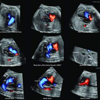
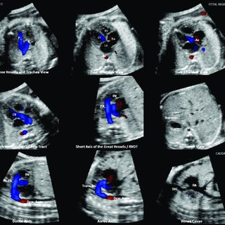
Fetal echocardiography is an ultrasound examination of the fetal heart, which includes fetal anatomic ultrasound planes of the upper abdomen, the 4 chamber view, the 5 chamber view, the short axis view, the 3-vessel-trachea , and if needed, longitudinal planes of the aortic arch, the ductal arch, and the systemic veins. Improved diagnostic accuracy of fetal echocardiography is attained by the application of gray scale and color Doppler ultrasound in order to demonstrate blood flow in the cardiac chambers and great vessels. While the atria, ventricles, and atrioventricular valves are simultaneously visualized in the 4-chamber-view, the great vessels are assessed in the left and right outflow tracts thus demonstrating their origin, course, and spatial relationships. Documentation of the fetal echocardiography examination is required and includes storing of still images and movie clips of cardiac anatomy. Review of the fetal echocardiography study at a later time is thus limited to the movie clips and still image obtained by the examiner.
The introduction of 3- and 4-dimensional (3D/4D) ultrasound, especially the spatial and temporal image correlation (STIC) about 15 years ago, added several advantages to fetal echocardiography including the ability to extract high number of ultrasound planes from 3D/4D volumes. Incorporating 3D/4D in fetal echocardiography has been used to display surface mode images with 2-dimensional (2D) and color Doppler, to extract single or multiple planes in tomographic view , thus enabling the offline examination of the heart, a technique used in telemedicine. Algorithms have been developed on how to extract planes out of a 3D/4D volume; an approach helpful in teaching fetal echocardiography, especially the outflow tracts. Furthermore, 3D/4D allows for volume calculations of cardiac chambers, which is used to accurately assess cardiac volumes in systole and diastole . In the past several years, novel applications of 3D/4D in fetal echocardiography emerged primarily in 3 different aspects: Color Doppler rendering, the matrix probe with biplane, or X-plane visualization and advances in the field of automation of the fetal heart.
Color flow Doppler, power Doppler, and bidirectional power Doppler can be combined with the STIC tool to acquire volumes including 2D and color information throughout the cardiac cycle. The image display can be the tomographic, orthogonal, or the 3D volume rendering mode. The information displayed in volume rendering can include either the color information alone, the gray-scale information alone, or a combination of both, termed “glass body” mode. A special software using a light source can in addition emphasize the depth effect of the blood flow and the surrounding structures and provides an enhanced spatial impression (Fig. 1).
We are a private company, one of the leading Laboratory & Medical devices, consumables and equipment distributors in the region with more than 35 years of history built with a mixture of boldness, innovation and visionary spirit that helped modeling our evolution and the evolution of the laboratory & medical sector itself.