
CrystalLive™ 3D
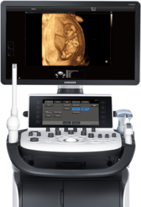
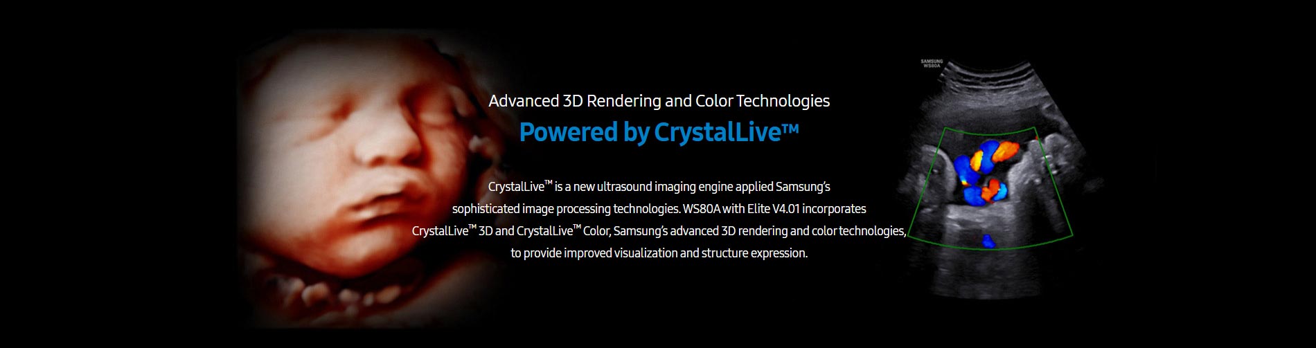
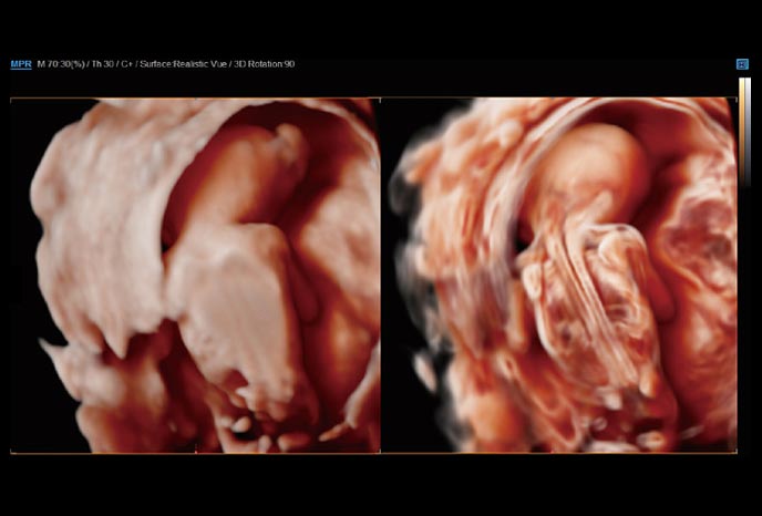
High Definition Volume Imaging™ (HDVI™) is a volume rendering technology that improves visualization of edges and small structures in volume data. HDVI™ provides upgraded marginal expression and image saturation with dedicated image sets for 3D/4D.
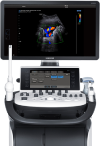
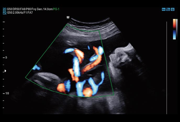
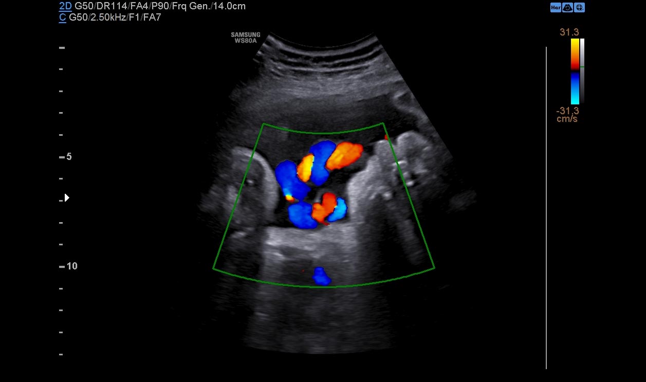
CrystalLive™ Color
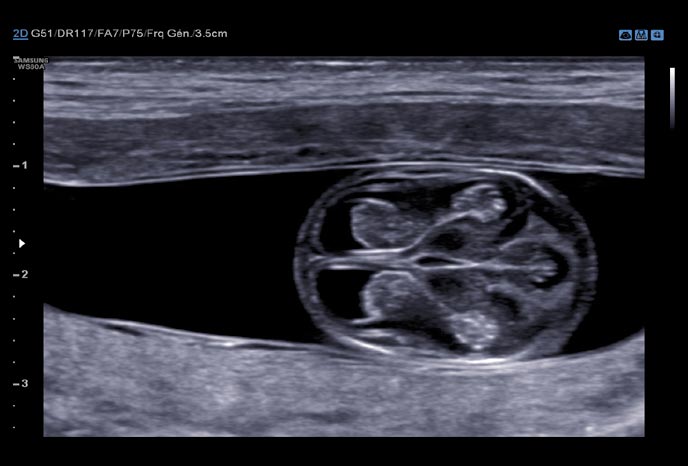
CrystalLive™ Color, Samsung’s advanced blood flow imaging technologies in color mode, enhances the visualization and hemodynamics of the blood flow.
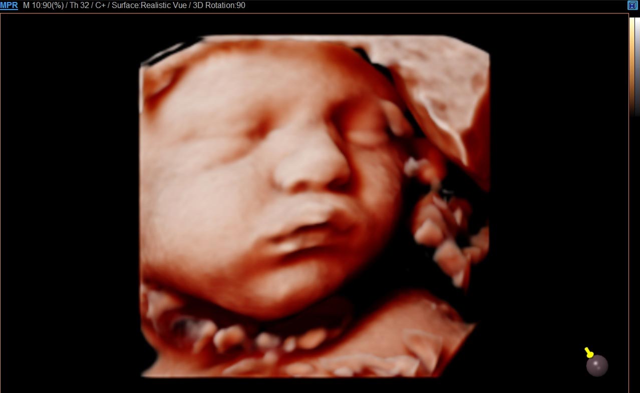
CrystalLive™ 3D
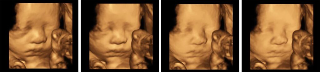
Margin enhancement with HDVI™
High Definition Volume Imaging™ (HDVI™) is a volume rendering technology that improves visualization of edges and small structures in volume data. HDVI™ provides upgraded marginal expression and image saturation with dedicated image sets for 3D/4D.
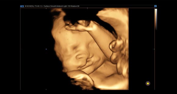
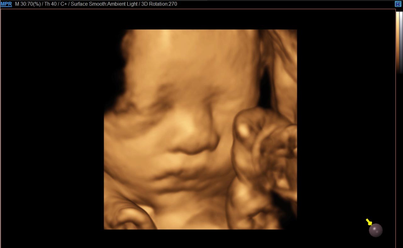
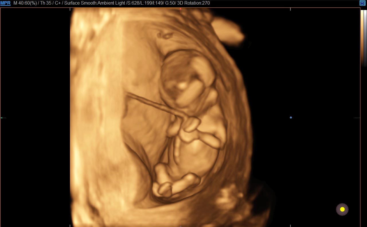
Improved structural description with AmbientLight
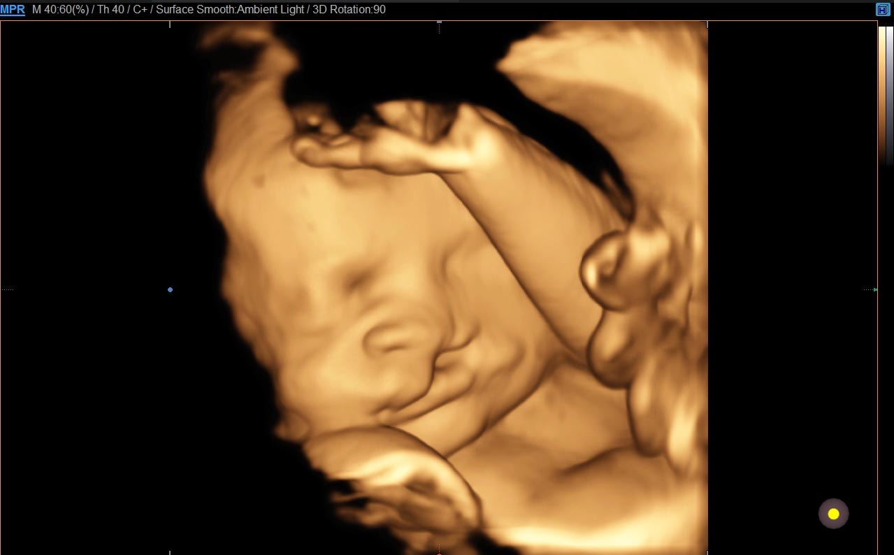
Creating intricately graduated shadows, AmbientLight improves depth expression of the surface. This 3D rendering feature is especially useful to see a fetal face or hands in detail.
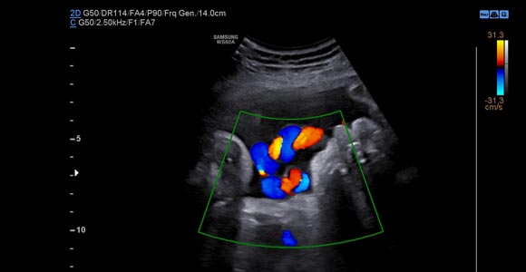
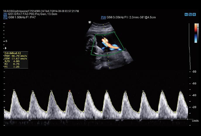
CrystalLive™ Color
CrystalLive™ Color, Samsung’s advanced blood flow imaging technologies in color mode, enhances the visualization and hemodynamics of the blood flow.
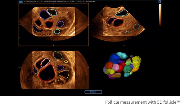
5D Follicle™ (Follicle measurement)
5D Follicle™ identifies and measures multiple ovarian follicles for rapid assessment of follicular size and status during gynecology examinations.
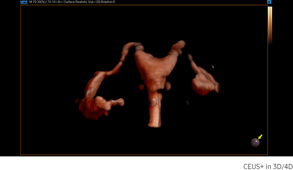
CEUS+
CEUS+ can be used in 3D/4D for effective examination for patency of the fallopian tube and morphology of uterus and endometrium.
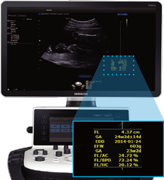
BiometryAssist™(Fetal biometry estimation)
A semi-automatic technology for biometric measurement,BiometryAssistant™, enables users to measure the growthof the fetus more quickly and with greater accuracy whilemaintaining exam consistency.
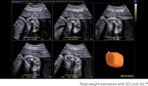
5D Limb Vol.™ (Fetal weight estimation)
5D Limb Vol™. is a semi-automated tool to quickly and accurately measure upper arm or thigh volumes from 3 simple seed points on a single volume data set. These measurements can then be used to calculate an accurate estimation of fetal weight as well as provide additional information regarding fetal nutritional status.
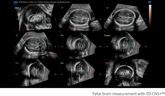
5D CNS+™ (Fetal brain measurement)
5D CNS+™ uses intelligent navigation to provide 6 measurements from 3 transverse views of the fetal brain to enhance measurement reproducibility and streamline workflow.It includes axial, sagittal and coronal views with 9 planes.
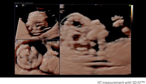
5D NT™ (Nuchal translucency measurement)
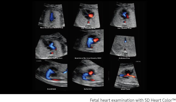
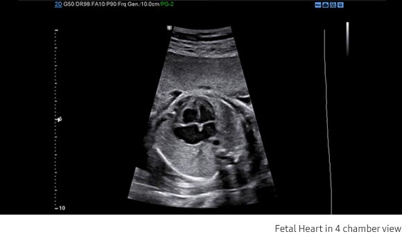
5D Heart Color™ (Fetal heart examination)
5D Heart Color™ uses STIC volume datasets.Color Doppler sonography is demonstrated in 9 standard fetalechocardiography views in a single display.
RealisticVue™
RealisticVue™ displays high resolution 3D anatomy with exceptional detail and realistic depth perception.User selectable light source direction creates intricately graduated shadows forbetter defined anatomical structures.
CrystalVue™
CrystalVue™ is an advanced volume rendering technology that enhances visualization of both internaland external structures in a single rendered image using a unique combination of intensity,gradient and position.
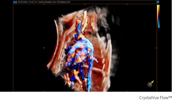
CrystalVue Flow™
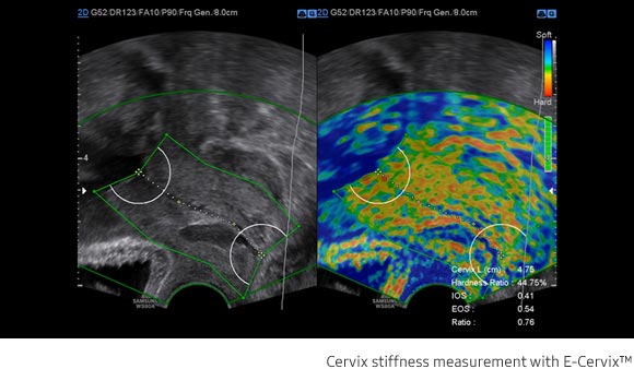
E-Cervix™ (ElastoScan™ for Cervix)
E-cervix™ shows the strain ratio between the internal and external orifices of the uterus using the vibrations caused by the natural internal movements.* This technology could increase reproducibility and reduce inter-observer variance using the sum of 50 elastographic images acquired over 3.5 seconds.
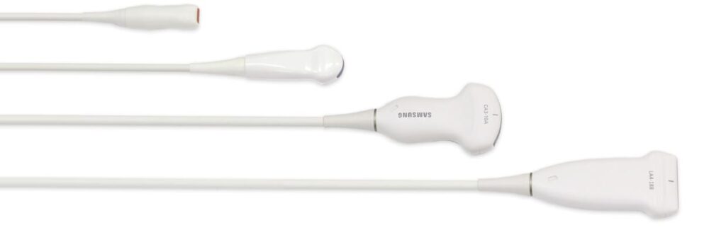
Neonatal / Pediatric transducers
Highly advanced transducers allow for excellent detailed resolutionand more efficient scanning.
User-friendly design
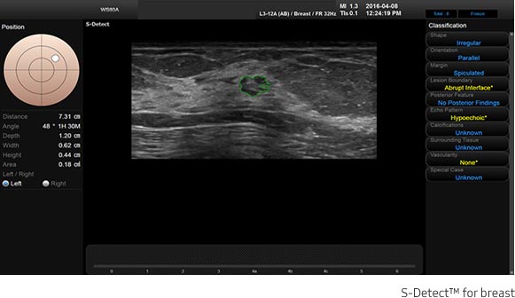
S-Detect™ (S-Detect™ for breast)
S-Detect™ for Breast helps standardize reporting and classification of suspicious breast lesions by incorporating BIRADS ® ATLAS* (Breast Imaging-Reporting and Data System, Atlas) into the tool. When the user selects a region of interest, S-Detect™ for Breast automatically defines the lesion boundaries, provides lexicon classification options and images export for an enhanced and streamlined workflow.
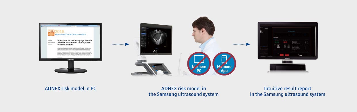
IOTA-ADNEX(Ovarian tumor classification)
Samsung has adopted the ADNEX* model for classifying ovarian tumors as proposed by the IOTA** group and named it IOTA-ADNEXNow all ultrasound procedures, from initial scan to the final report, can be carried out using the same system.It even fills in 2 ultrasound predictors*** automatically, as soon as they are measured,and provides a report that classifies the results according to the ADNEX model 5-level classificationfor ovarian tumors to help users make the appropriate decisionsfor managing the patient’s condition.
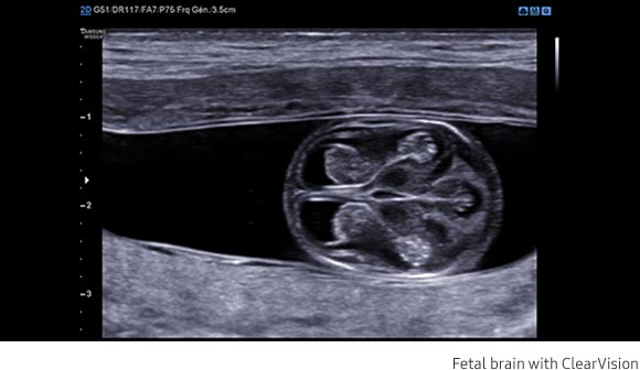
ClearVision
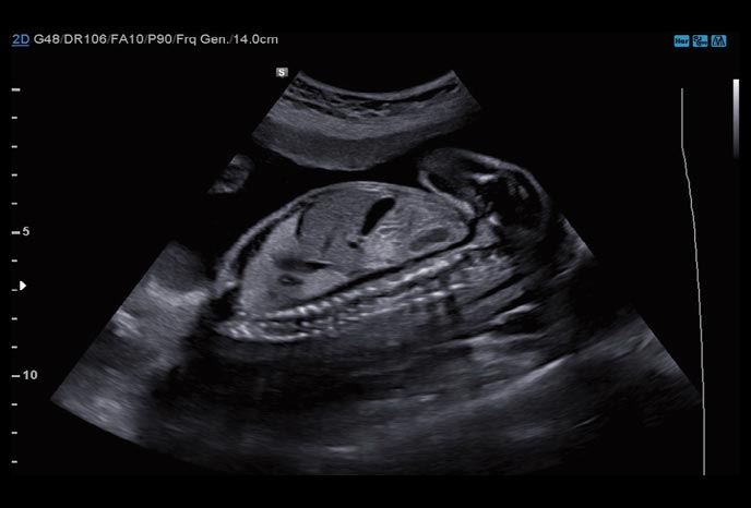
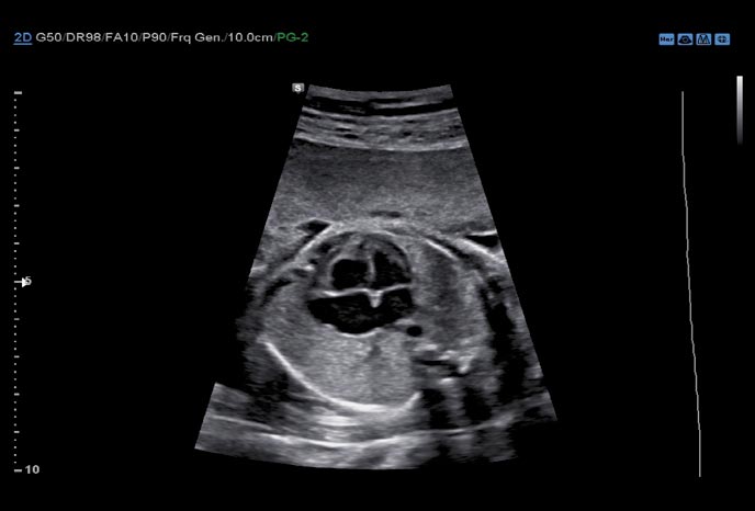
The noise reduction filter improves edge enhancement and creates sharper 2D images for optimal diagnostic performance. The integration of specialized Samsung technology results in a notable improvement in image quality.
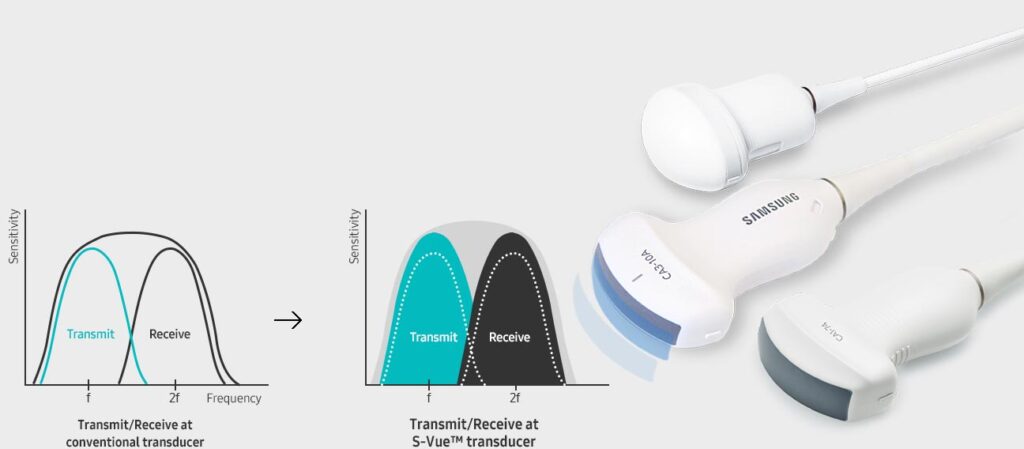
S-Harmonic™
This new harmonic technology improves image clarity, near to far.Reducing signal noise, S-Harmonic provides more uniform ultrasound images.
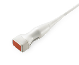
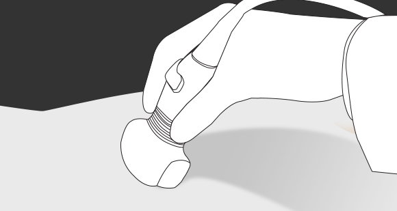
In addition, the ergonomically designed S-Vue transducer fits well in the hand and is easy to handle.
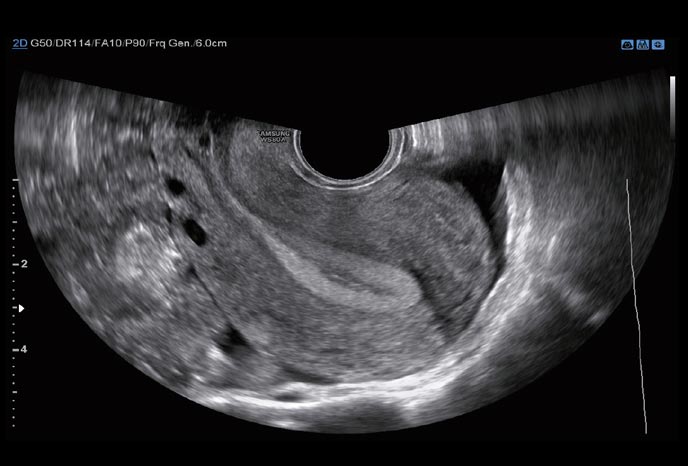
Wide angle endocavity transducer
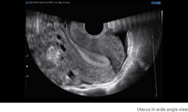
The new wide angle endocavity transducer (E3-12A) offers a field-of-view up to 210° allowing greater visualization of pelvic anatomy. It is often possible to visualize the entire cervix and uterus in normal anatomy as well as viewing left-right symmetry in the transverse plane.
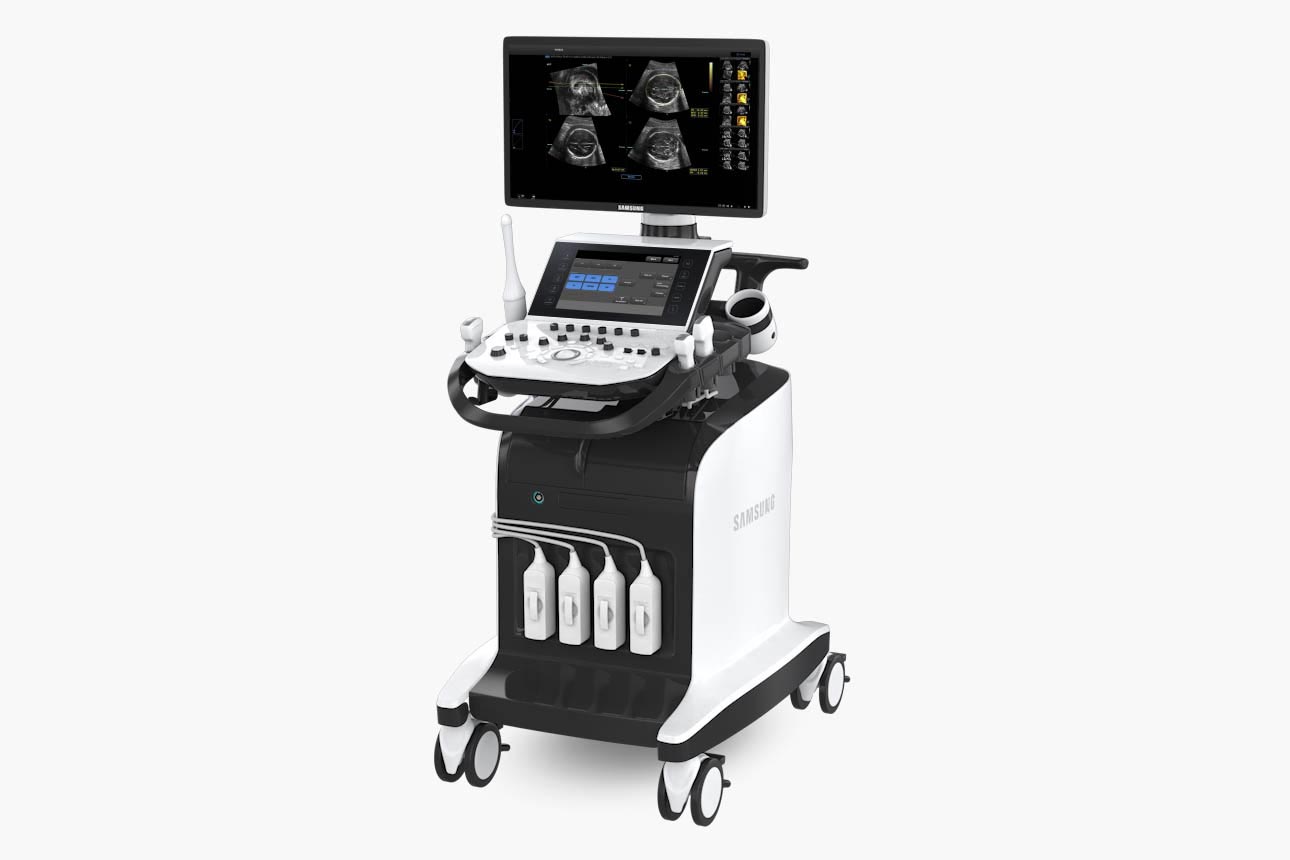
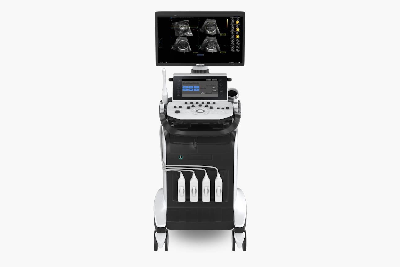
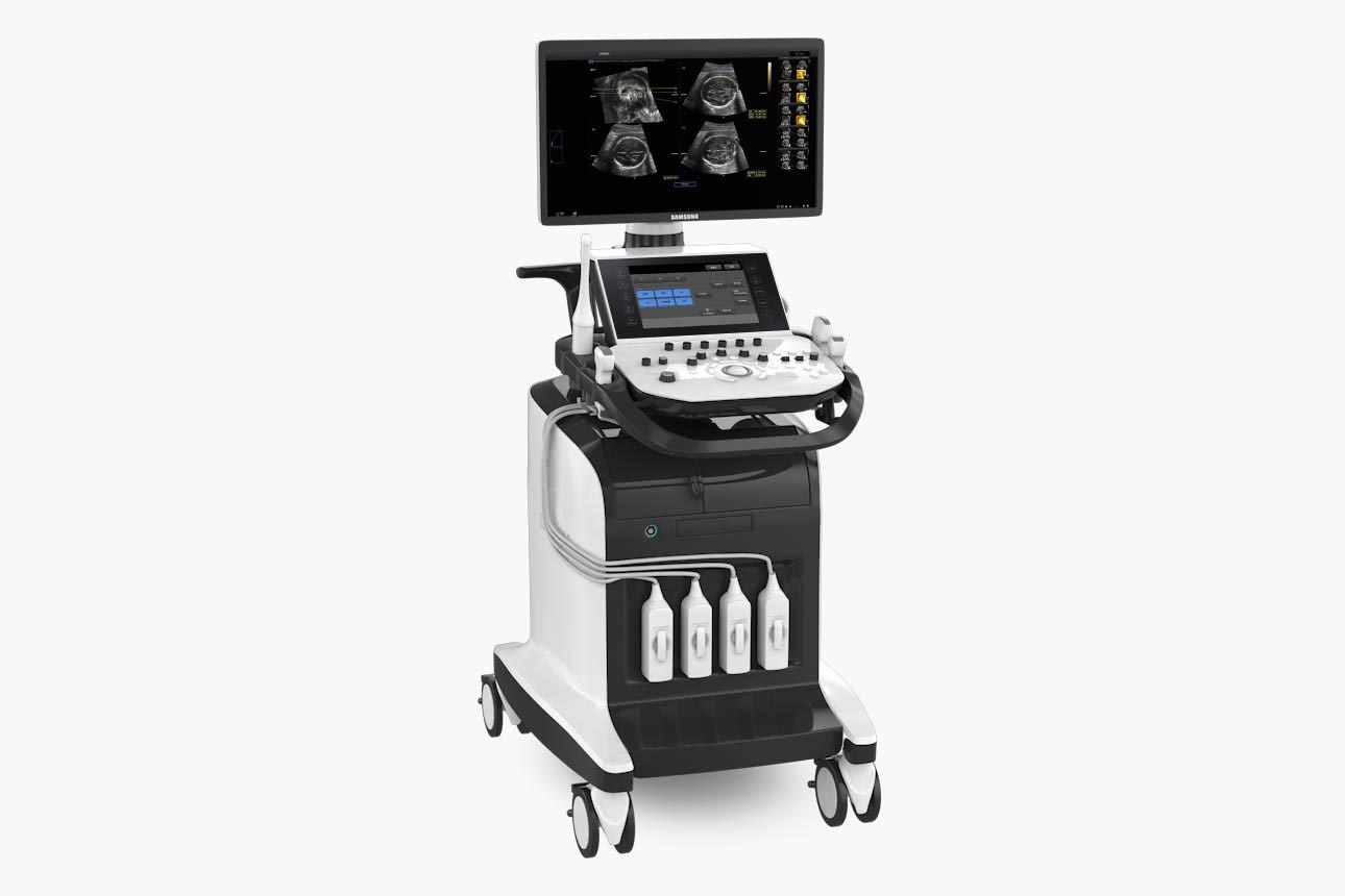
The WS80A with Elite features a 23.8-inch full HD LED display, delivering excellent contrast resolution, image clarity and vibrant color in any lighting condition.
Two-level adjustable gel warmer maintains ultrasound gel at a comfortable temperature from 30°C to 39°C, which comes as a default feature on WS80A with Elite.
Smooth up and down lift allows you to adjust the system to the your preferred height without straining.
The 10.1-inch touch screen is highly sensitive, allowing for easy input to minimize users’ stress from repetitively pressing buttons.
Users can arrange the Transducer cables neatly on the two hangers on either side of the system.
The WS80A with Elite features endocavity transducer holders for convenience when performing gynecological scanning and discrete storage when not in use.
SonoSync™*For real-time image sharing solution
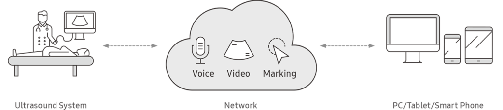
SonoSync™ is a real-time image sharing solution that allows collaborative communication for care guide and training between doctors and sonographers. In addition, voice chatting and real-time marking function are provided for efficient communication, and the MultiVue function is included to monitor multiple ultrasound images on a single screen.

Application: Abdomen, Obstetrics, Gynecology
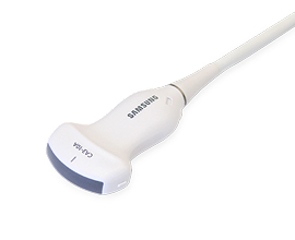
Application: Abdomen, Obstetrics, Gynecology, Pediatric
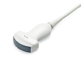
Application: Abdomen, Obstetrics, Gynecology, Musculoskeletal
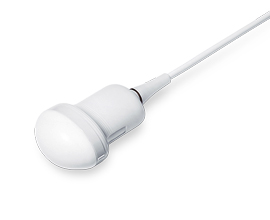
Application: Abdomen, Obstetrics, Vascular
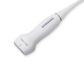
Application: Abdomen, Cardiac, TCD
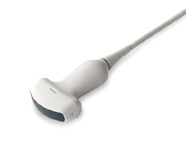
Curved array transducers

Application: Abdomen, Obstetrics, Gynecology
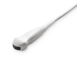
Application: Pediatric, Vascular
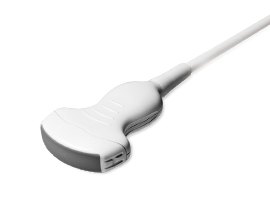
Application: Abdomen, Obstetrics, Gynecology
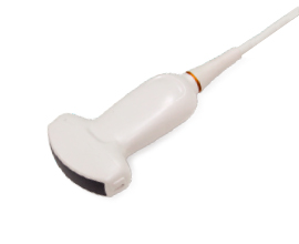
Application: Abdomen, Obstetrics, Gynecology
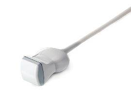
Volume transducers

Application: Musculoskeletal, Small parts, Vascular
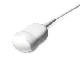
Application: Abdomen, Obstetrics, Gynecology
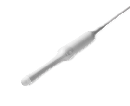
Application: Obstetrics, Gynecology, Urology
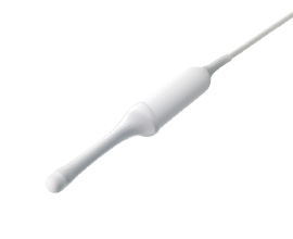
Application: Obstetrics, Gynecology, Urology
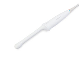
Application: Obstetrics, Gynecology, Urology
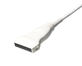
Linear array transducers

Application: Small parts, Vascular, Abdomen, Musculoskeletal
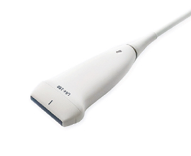
Application: Small parts, Vascular, Musculoskeletal
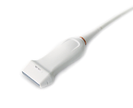
Application: Small parts, Vascular, Musculoskeletal
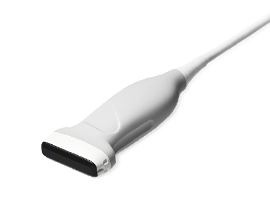
Application: Small parts, Vascular, Musculoskeletal
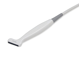
Application: Musculoskeletal
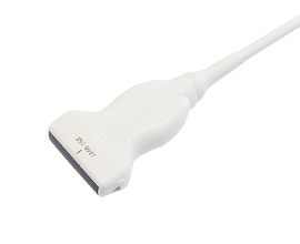
Application: Small parts
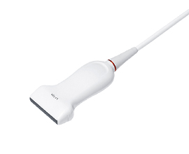
Application: Small parts, Vascular, Obstetrics, Musculoskeletal, Abdomen
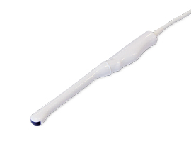
Endocavity transducers

Application: Obstetrics, Gynecology, Urology
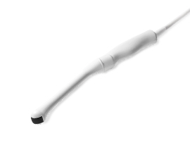
Application: Obstetrics, Gynecology, Urology
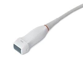
Phased array transducers

Application: Abdomen, Cardiac, TCD

Application: Abdomen, Cardiac, TCD
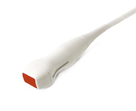
Application: Abdomen, Pediatric, TCD
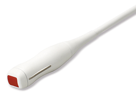
Application: Cardiac, Pediatric
We are a private company, one of the leading Laboratory & Medical devices, consumables and equipment distributors in the region with more than 35 years of history built with a mixture of boldness, innovation and visionary spirit that helped modeling our evolution and the evolution of the laboratory & medical sector itself.