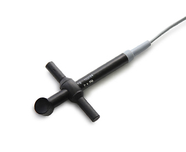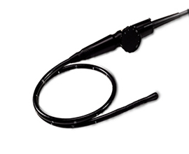
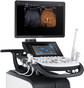
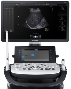

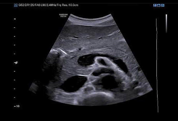
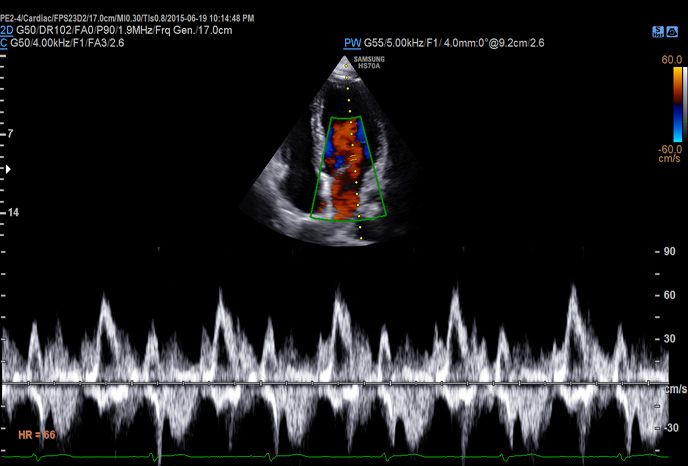
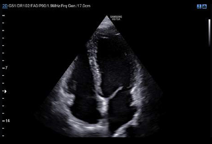
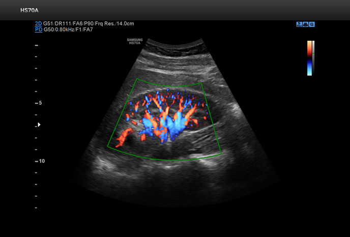
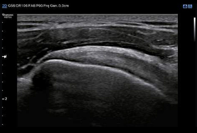
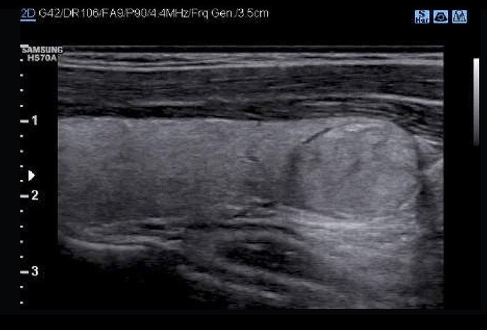
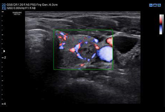
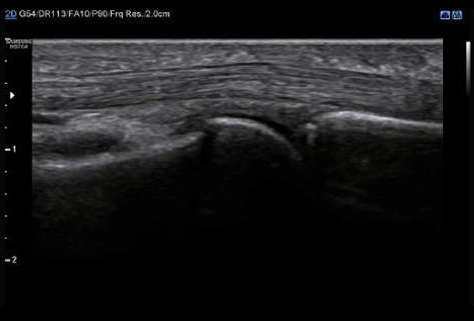
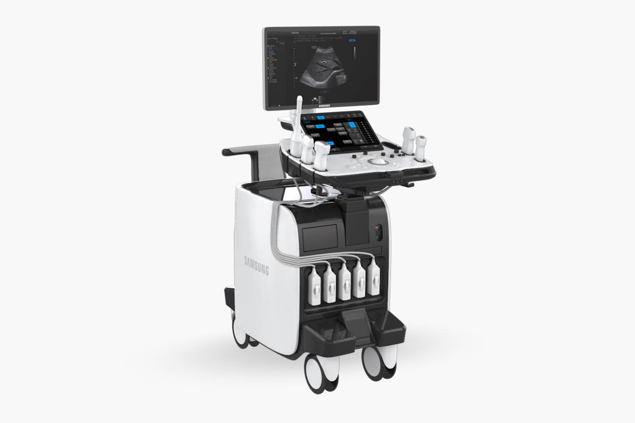
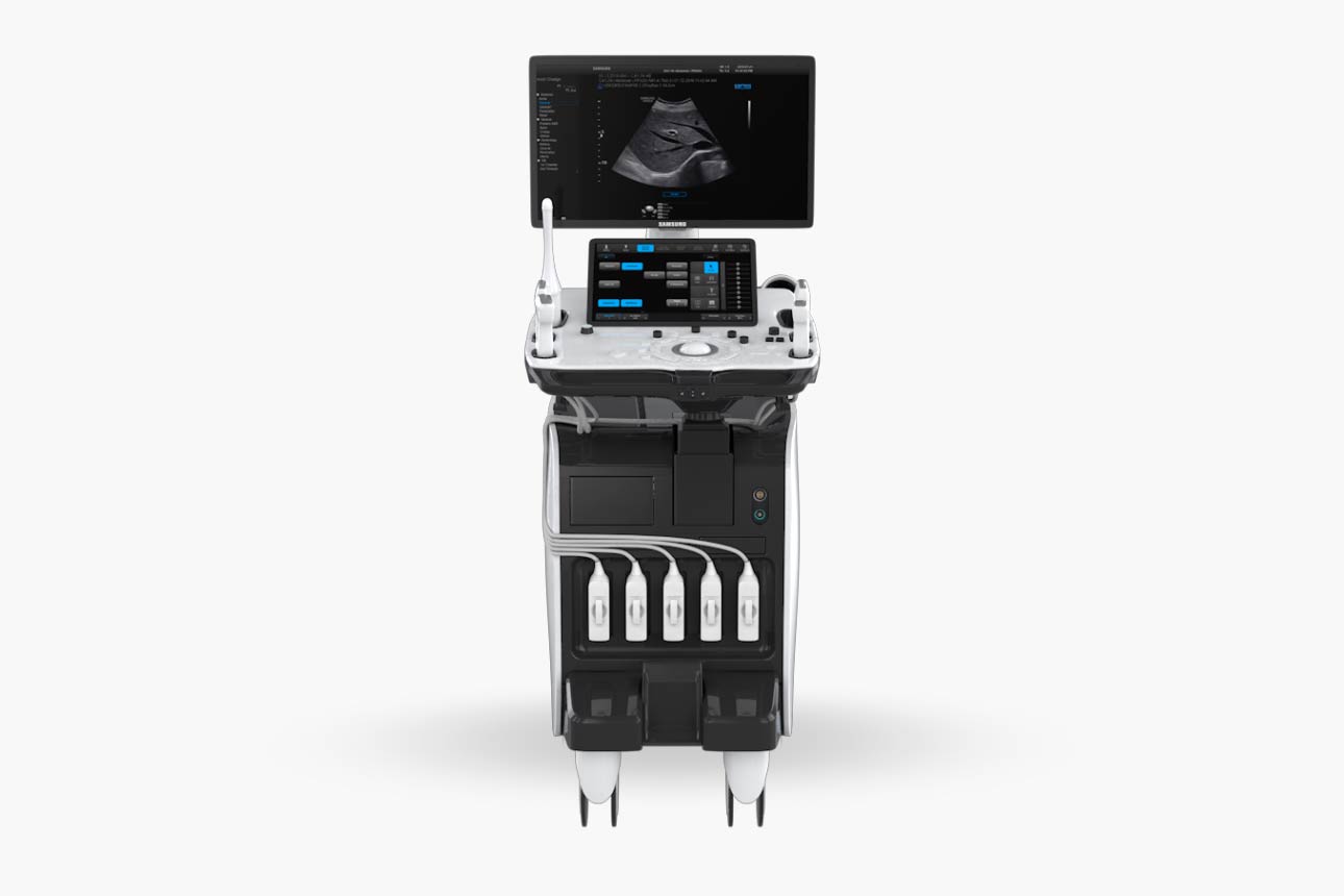
HS70A with Prime

Easier and quantitative Measurement

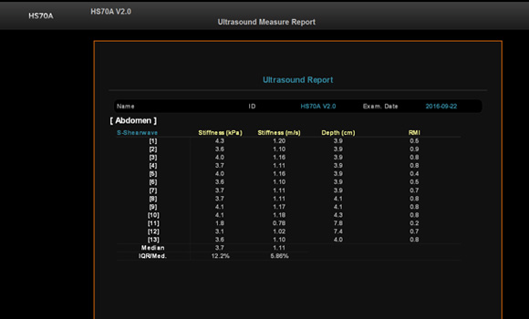
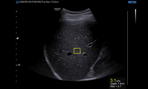
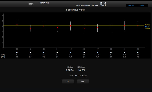
S-Shearwave detects the velocity of the shearwave  propagated through the targeted lesion and displays the numerical measurement of stiffness In kPa or m/s together with a Reliable Measurement Index (RMI)*. 

HS70

Visualization of more information

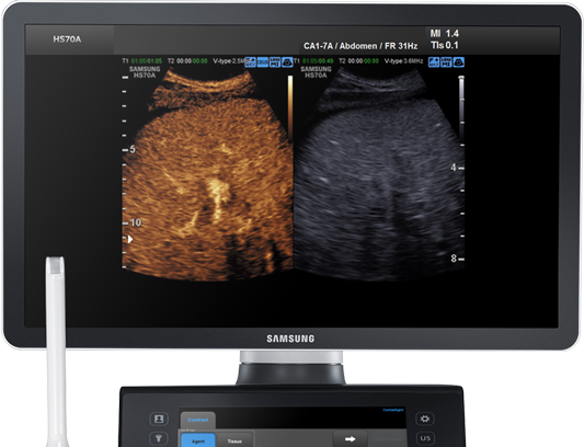
CEUS+ technology uses the unique properties of ultrasound contrast agents. When stimulated with low MI frequencies, the oscillating microbubbles reflect both basic frequencies and harmonic signals.

Visible lesions, easier examination

CEUS+

CEUS+ technology uses the unique properties of ultrasound contrast agents.When stimulated with low MI frequencies, the oscillating microbubbles reflect both basic frequencies and harmonic signals.
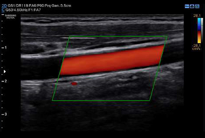
In addition, Samsung’s latest technologies provide a clear visualization ofvessels and blood flow so that you can form an informed, reliable diagnosis with confidence.

S-Shearwave

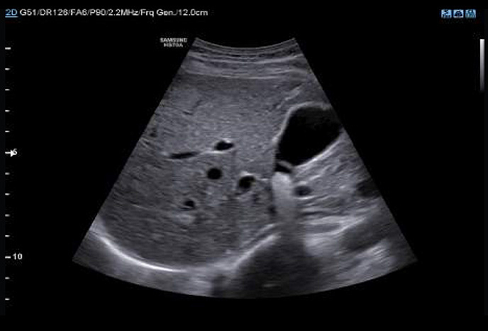
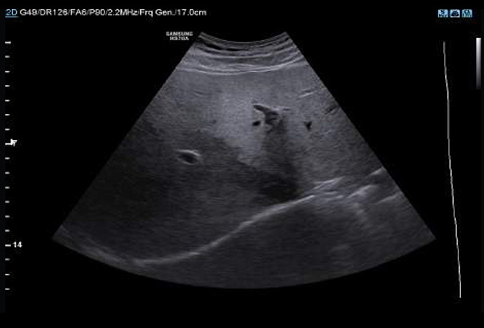
S-Shearwave detects the velocity of the shearwave propagated through the targeted lesion and displays the numerical measurement of stiffness In kPa or m/s together with a Reliable Measurement Index (RMI)*. S-Shearwave has the potential to reduce the number of conventional liver biopsies by providing quantitative tissue characteristic information.

Advanced measurement tools

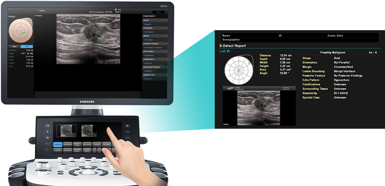
S-Detect for Breast

By simply clicking the suspected area, S-Detect™ for Breast draws the lesion area andprovides the characteristics of the lesion and a recommendation on whetherthe lesion is benign or malignant.

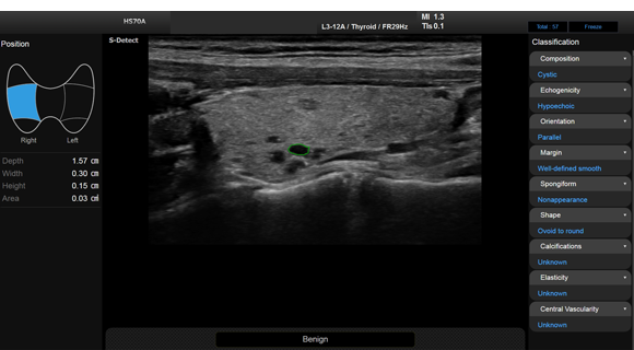
S-Detect for Thyroid

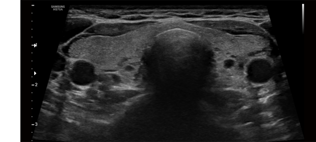
S-Detect™ for Thyroid uses the advanced technology based on *K-TIRADS, RUSS and ATA draft in detecting and classifying suspicious thyroid lesions semi-automatically. This state-of-the art technology helps you diagnose your patients with confidence and ease, providing accurate, consistent results and an automatic reporting feature.

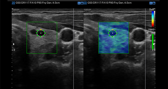
E-Thyroid

E-Thyroid™ uses pulsations from the adjacent Carotid Artery and provides an assessment of thyroid lesions.

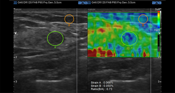
E-Strain

E-Strain enables quick and easy calculation of the strain ratio between two regions of interest in day-to-day procedures such as breast, prostate or gynecological examinations.

Preventive actions

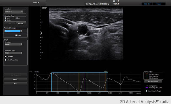
Arterial Analysis

Arterial Analysis detects functional changes of vessels, providing measurement values such as the stiffness and intima-media thickness. Since the functional changes occur before morphological changes, this technology supports the early detection of cardiovascular diseases.

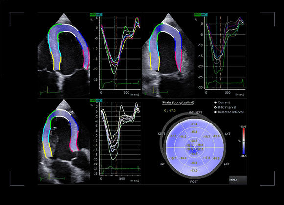
Strain+

Strain quantitativelydisplays a Bull’s Eye which shows leftventricular motion and dyssynchrony at a glance.

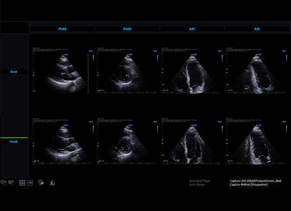
Stress Echo

The Stress Echo package includes wall motion scoring and reporting.
It includes exercise Stress Echo, pharmacologic Stress Echo, diastolic Stress Echo and free programmable Stress Echo.

Image clarity

S-Vision imaging engine

The S-Vision Beamformer receives returning signals through a sophisticated digital filtering system resulting in reduced side lobes, less noise and artifact.

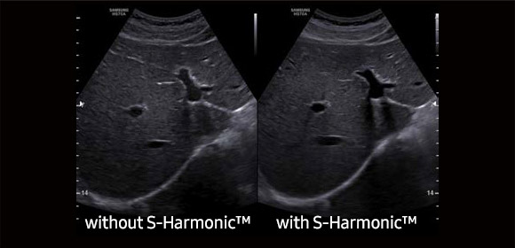
S-Harmonic

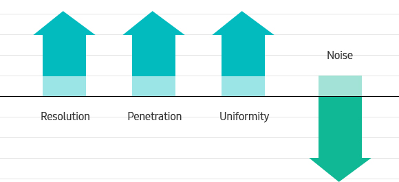
This new harmonic technology makes a clearer image – near to far.
Reducing signal noise,S-Harmonic provides more uniform ultrasound images.


ClearVision

ClearVision offers speckle reduction, edge enhancement and contrast enhancement for clear and natural images. In addition, ClearVision provides application-specific optimization in live scan mode.

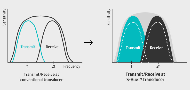
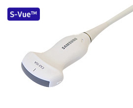
Imaging becomes Even Better with S-Vue™ transducer

(Single Crystal Technology)

Employing an innovative crystal design, The S-Vue™ transducer provides more efficient piezoelectric properties, resulting in wider bandwidths for increased depth penetration and higher quality resolution on even the most challenging patient.

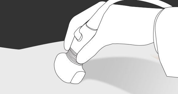
In addition, the ergonomically designedS-Vue transducer fits well in the hand andis easy to handle.

Quick Preset

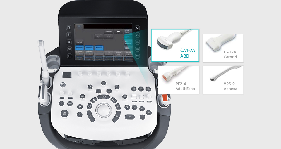
Quick Preset shows the four connected transducers and, for each of them, the most frequently used image settings. With one touch, the desired transducer.

EZ-Exam+

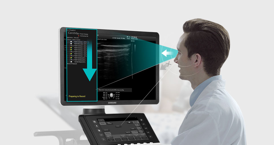
EZ-Exam+™ transforms multiple imaging steps into a streamlined process. It stores optimized and most frequently used protocols in a button.

Advanced QuickScan

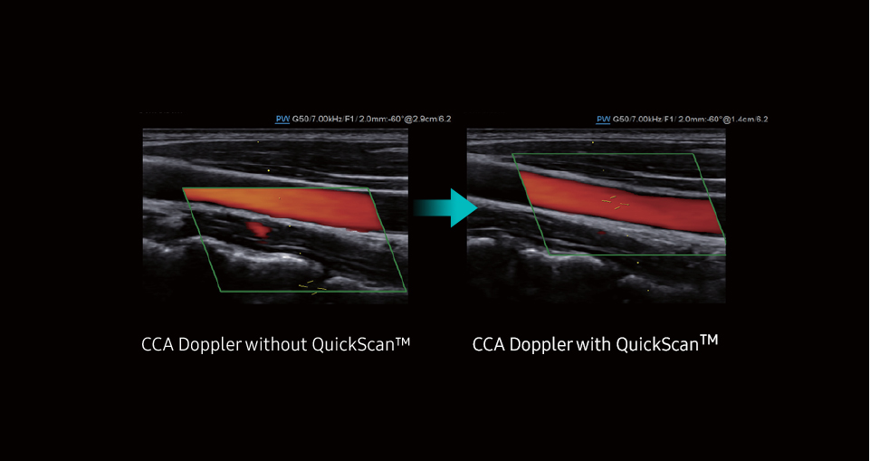
Advanced QuickScan™ technology provides intuitive optimization of gray scale and Doppler parameters. One touch of the QuickScan™ button elevates efficiency and workflow by adjusting functions including color gain and color box location.

Ergonomic Design

Created by potrace 1.16, written by Peter Selinger 2001-2019

Silent operation

This exceptionally quiet device allows physical exams to be performed, including auscultation, while the ultrasound system is turned on.

Created by potrace 1.16, written by Peter Selinger 2001-2019

23-inch LED Back light display

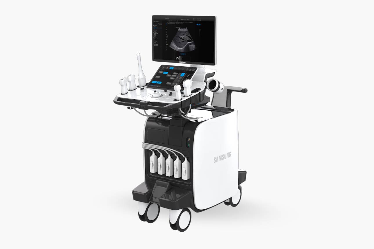
To enhance the image, the HS70A with Prime features a 23-inch full high-definition (FHD) LED display, delivering superior image contrast on a large ultrasound display.

Created by potrace 1.16, written by Peter Selinger 2001-2019

Gel warmer

For operator convenience, a gel warmer can be installed on both sides of the control panel.

Created by potrace 1.16, written by Peter Selinger 2001-2019

Silent operation

This exceptionally quiet device allows physical exams to be performed, including auscultation, while the ultrasound system is turned on.

Created by potrace 1.16, written by Peter Selinger 2001-2019

10.1-inch touch screen

The 10.1-inch touch screen is exceptionally sensitive and makes operating the ultrasound system smartly efficient.

Created by potrace 1.16, written by Peter Selinger 2001-2019

User-friendly console design

Customizable U and P keys allow users to create a workflow tailored to their needs. The console also can be adjusted up, down, left and right so each user is ensured the optimal location.

Clinical Images

Showroom

Transducers

Curved array transducers

CA3-10A

Application: Abdomen, Obstetrics, Gynecology, Pediatric, Vascular
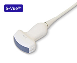
CA2-9A

Application: Abdomen, Obstetrics, Gynecology
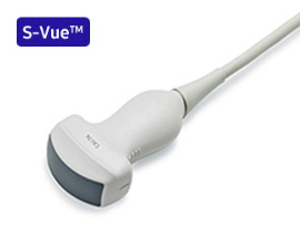
CA1-7A

Application: Abdomen, Obstetrics, Gynecology, Musculoskeletal
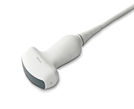
CA2-8A

Application: Abdomen, Obstetrics, Gynecology
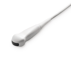
CF4-9

Application: Pediatric, Vascular

Linear array transducers

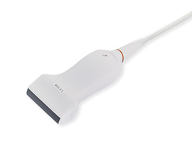
L3-12A

Application: Small parts, Vascular, Musculoskeletal, Obstetrics, Abdomen
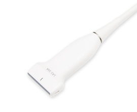
LA3-16A

Application: Small parts, Vascular, Musculoskeletal
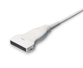
LA2-9A

Application: Small parts, Vascular, Musculoskeletal, Abdomen
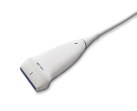
LA4-18B

Application: Small parts, Vascular, Musculoskeletal
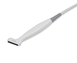
LA3-16AI

Application: Musculoskeletal, Intra-operative
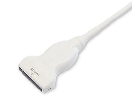
LM4-15B

Application: Small parts, Vascular, Musculoskeletal, Abdomen
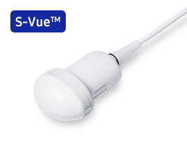
Volume transducers

CV1-8A

Application: Abdomen, Obstetrics, Gynecology
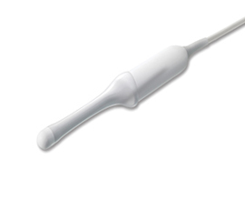
V5-9

Application: Obstetrics, Gynecology, Urology
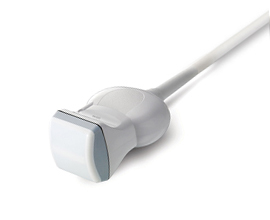
LV3-14A

Application: Musculoskeletal, Small parts, Vascular
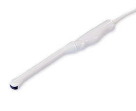
Endo-cavity transducers

EA2-11B

Application: Obstetrics, Gynecology, Urology
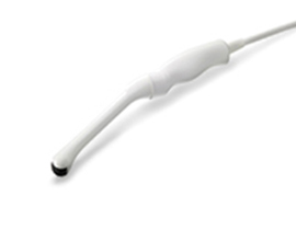
VR5-9

Application: Obstetrics, Gynecology, Urology
Phased array transducers

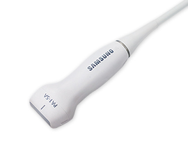
PA1-5A

Application: Cardiac, Pediatric

PA4-12B

Application: Cardiac, Pediatric

PE2-4

Application: Abdomen, Cardiac, TCD

PA3-8B

Application: Cardiac, Pediatric, Abdomen
CW transducers

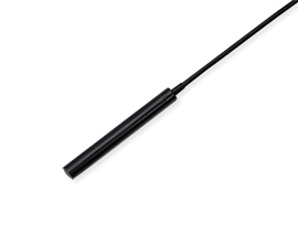
DP8B

Application: Cardiac, Vascular

DP2B

Application: Cardiac

TEE transducer

MMPT3-7

Application: Cardiac